From Mud to Microscope:
Sediment Processing Alexandrium catanella cysts
Anushka Rajagopalan
6/27/23
Alexandrium catenella is a neurotoxin-producing dinoflagellate that can proliferate into a Harmful Algal Bloom (HAB). This species begins its life cycle as a resting cyst on the seafloor which then germinates into cells that can photosynthesize and create toxic blooms. Many researchers are studying the effects of A. catenella on human and ecosystem health. For example, in my lab, one of my roles is to analyze sediment samples from off the coast of Alaska to observe if certain environmental factors impact cyst abundance and bloom initiation.
But how do these samples get processed in the first place?
This blog highlights the process of staining A. catenella cysts from sediment samples collected in the field to determine their abundance across a specific geographical area. This specific protocol is used by the Donald Anderson Laboratory at the Woods Hole Oceanographic Institution. The sediments used in this protocol were collected during an Alaskan Arctic Cruise in 2022. Each sample was labeled with its specific location/transect. Each sample has 2 ccs that are measured out and placed into a 50 mL tube with an equal amount of cold filtered seawater to rinse the sediment. This is probably the muddiest part – gloves are essential to make it through the day!
The rinsed sediment is sonicated with a Branson 250 Sonifier, a high-frequency tool that uses ultrasonic energy to agitate, mix, and break up clumps of sediment for 60 seconds at 40% amplitude. It also kind of sounds like you’re at the dentist at this point.


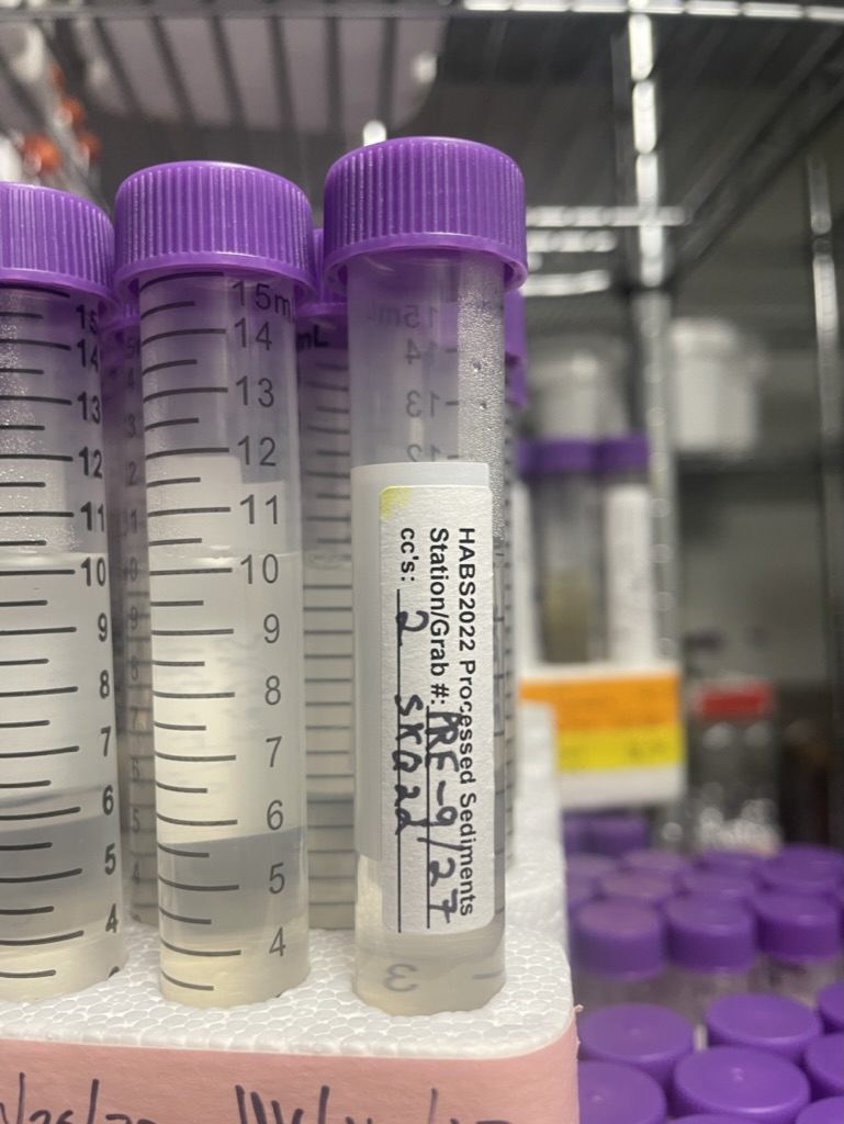
The sample is then sieved sequentially with an 80 µm and 20 µm Nitex screen to isolate the size class of cysts. Sometimes the work can get slowed down if there are huge clumps – definitely important to be patient during this. After sieving, the sample is transferred to a 15 mL centrifuge tube and 0.5 mL of 100% formalin is added to preserve any proteins and cellular organelles left in the sample. The next day, we extract these samples with MeOH and use a dye called Primulin – this will allow the cysts to fluoresce and can be counted with microscopy. The volumes used for this process can change based on specific protocols.
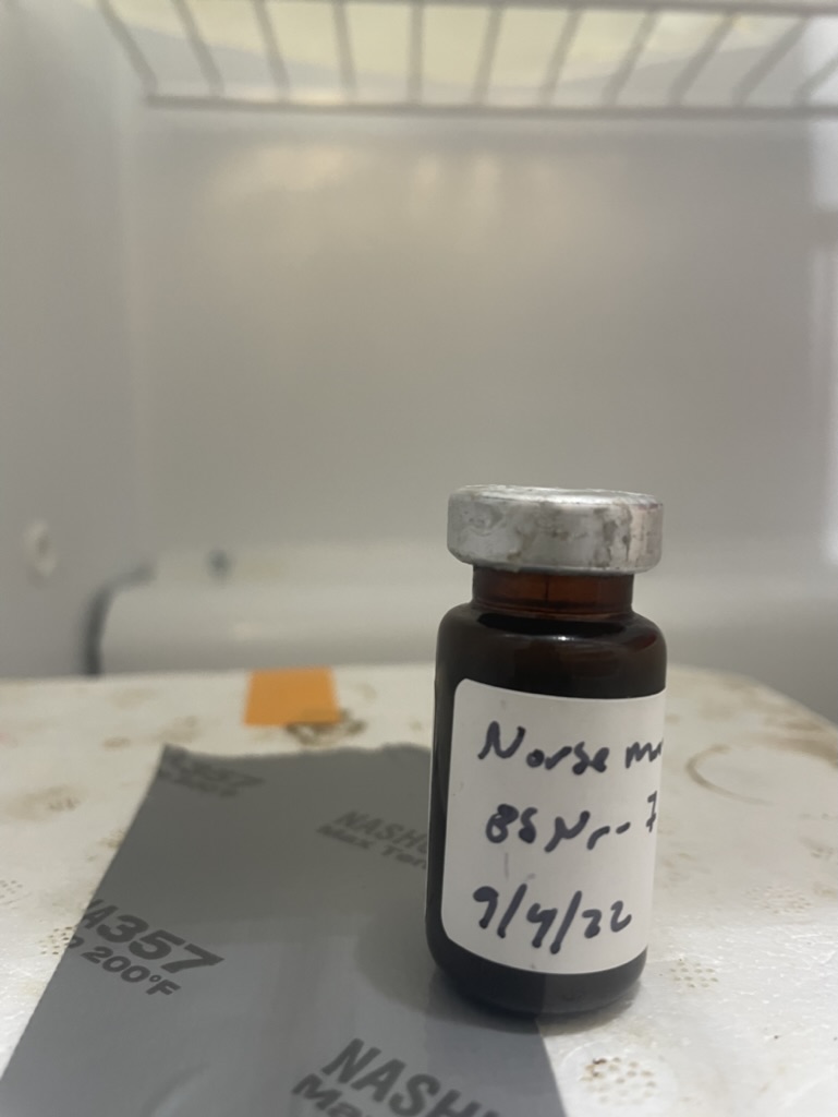
Sediment samples pre and post processing. Photo credit: Anushka Rajagopalan, WHOI
This is an important procedure that highlights the steps collecting data to study A. catenella cysts. The sediment processing described here is the first step in collecting cyst data that we can then relate to other environmental variables. Usually, processing these samples takes all day for weeks at a time – it can be tedious work, but ultimately greatly contributes to investigating toxic blooms in the Arctic.
Reference: Anderson DM, Fachon E, Pickart RS, Lin P, Fischer AD, Richlen ML, Uva V, Brosnahan ML, McRaven L, Bahr F, Lefebvre K, Grebmeier JM, Danielson SL, Lyu Y, Fukai Y. Evidence for massive and recurrent toxic blooms of Alexandrium catenella in the Alaskan Arctic. Proc Natl Acad Sci U S A. 2021 Oct 12;118(41):e2107387118. doi: 10.1073/pnas.2107387118. PMID: 34607950; PMCID: PMC8521661.