SIMS Basics
The SIMS (Secondary Ion Mass Spectrometry) is a spatially high resolution analytical technique that is being used for analysis of isotopic composition of elements in small volumes of solid samples. In contrast to an electron microprobe, which utilizes an electron beam to bombard a solid sample and analyses intensities of characteristic X-rays of chemical elements, a SIMS analytical system (ion microprobe) uses a finely focused beam of primary ions (usually these are Cs+, O+, O2+, O- and O2-) directed onto a sample to obtain mass spectra of the ionized particles ejected from the sample due to collision cascade reactions.
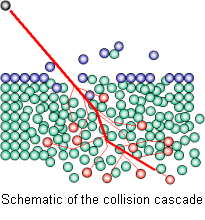
Schematic of the collision cascade.
In more detail, such interaction of primary ions with sample surface causes three major effects:
- Mixing of the upper layers of the sample that results in an amorphization of the material structure;
- Implantation of the primary ion beam atoms in the sample, and
- Ejection of particles (atoms or small molecules) from the sample surface which can be electrically neutral, as well as positively and negatively charged.
The charged particles of one polarity called "secondary ions" extracted and accelerated by electrostatic field form a secondary ion beam directed into a double focusing mass spectrometer where they are separated according to their energy and mass/charge ratio before being detected.
Nearly all elements of the periodic table (from H to U) can be quantitatively analyzed by SIMS technique. There are three basic modes of ion microprobe operation:
- Local analysis in a point. In this mode, a focused stationary primary beam is used to determine chemical (element concentrations) or isotopic compositions of the sample. The spot size ranges normally from a few to tens of microns and is selected for specific applications.
- Depth profiling. It is based on a phenomenon of etching a crater into the sample. Continuously collected secondary ions as the primary ion beam erodes the sample surface reveal compositional and isotopic changes with depth (i.e., by measuring signal intensity vs. time and then converting time into depth).
- Secondary ion imaging. The distribution of elements or isotopes can be obtained as an image of a given spatial resolution that could be reached of about ~1um over areas up to 500 μm2.