Gordon and Betty Moore Foundation Project Data
Microscale mechanistic linkages between the chemical and physical processes that contribute to marine organic matter degradation. 2015 - present.
Benjamin Van Mooy (WHOI) and Roman Stocker (ETH Zurich)
Particulate organic matter (POM) is ubiquitous in marine systems, and its role in the global carbon cycle is defined by microscale interactions between POM and microbes (Jackson, 1989; Azam, 1998; Jackson and Burd, 2002; Malfatti and Azam, 2009; Stocker, 2012). Microbial degradation drives the turnover of the entire reservoir of suspended POM in the surface ocean, which is composed primarily of living or recently dead plankton, on the timescale of every few weeks (IPCC, 2013). The suspended POM that escapes microbial degradation, forms aggregates (e.g., marine snow or fecal pellets), and sinks, is the primary vector of carbon from the surface ocean to the deep ocean (‘biological pump’). However, microbial degradation attenuates this flux as particles sink through the deep sea (Knauer et al., 1979; Martin, 1987; Van Mooy et al., 2002; Buesseler et al., 2007). Since the reservoir of carbon in the surface ocean is in near equilibrium with the CO2 in the atmosphere, the activity of microbes on sinking POM is a primary control on Earth’s climate (Kwon et al., 2009).
Owing to the importance of POM in the marine carbon cycle, POM degradation has been the focus of intense study since the dawn of modern oceanography, and yet this research has yielded very little mechanistic understanding of the microscale chemical and physical interactions underpinning POM degradation. Instead, most work has focused on characterizing POM composition and POM-microbe interactions at the bulk level. For example, bulk transformations in the chemical composition of sinking POM are characterized by a disappearance of readily hydrolysable organic matter, which contributes to the attenuation of the vertical POM flux (Wakeham et al., 1997; Armstrong et al., 2002; Van Mooy et al., 2002; Goutx et al., 2007). Similarly, it is understood that microbes physically interact with sinking POM by encountering particles by Brownian motion or motility, which contributes to dissolution and disaggregation of sinking POM and attenuates flux (Jackson, 1989; Burd et al., 2010). These are among the many bulk chemical and physical effects on POM flux attenuation that have found their way into simple particle export models, and yet the skill of these models is largely insufficient to obtain the predictive value required for inclusion in global climate and carbon biogeochemistry models.
We posit that developing an understanding of the mechanistic linkages between the microscale chemical and physical processes that contribute to POM degradation is critical to improve models of the marine carbon cycle.
We propose a pilot-scale study that couples Van Mooy’s recent work on understanding chemically mediated microscale connections between POM and microbes (Hmelo et al., 2011; Van Mooy et al., 2012; Edwards et al., 2015) with Stocker’s recent work on physical interactions between microbes and individual particles at the microscale (Seymour et al., 2010; Stocker, 2012).
Our goal for this proposed research is to establish relationships between the molecular signatures and microbial mechanisms of POM degradation at the scale of an individual particle. The long-term ambition is to be able to use observations of the molecular composition of sinking POM in the ocean to inform mechanistic models of particle flux attenuation. Our recent work has shown that the chemical composition of sinking POM directly influences flux attenuation through its effects on microbial interactions (Edwards et al., 2015), but we still lack the mechanistic context to establish predictive links to microscale physics behind these interactions.
Exploring the consequences of microbial communication bloom dynamics and nutrient cycling in the North Atlantic Ocean.
DYEatom Cruise
Enzyme hydrolysis rates from DYEatom cruise (R/V Pt. Sur; June 27 to July 5, 2013)<
Author: Bethanie Edwards
Posting Date: April 1, 2014
Excel file
Oxylipin and Lipid analysis
Fredricks / Van Mooy lab data filesOxylipin / lipid analysis
General information - all analyses
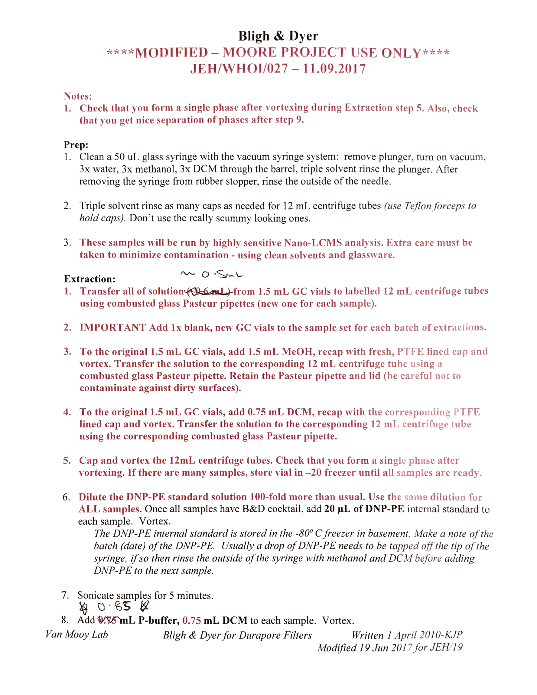
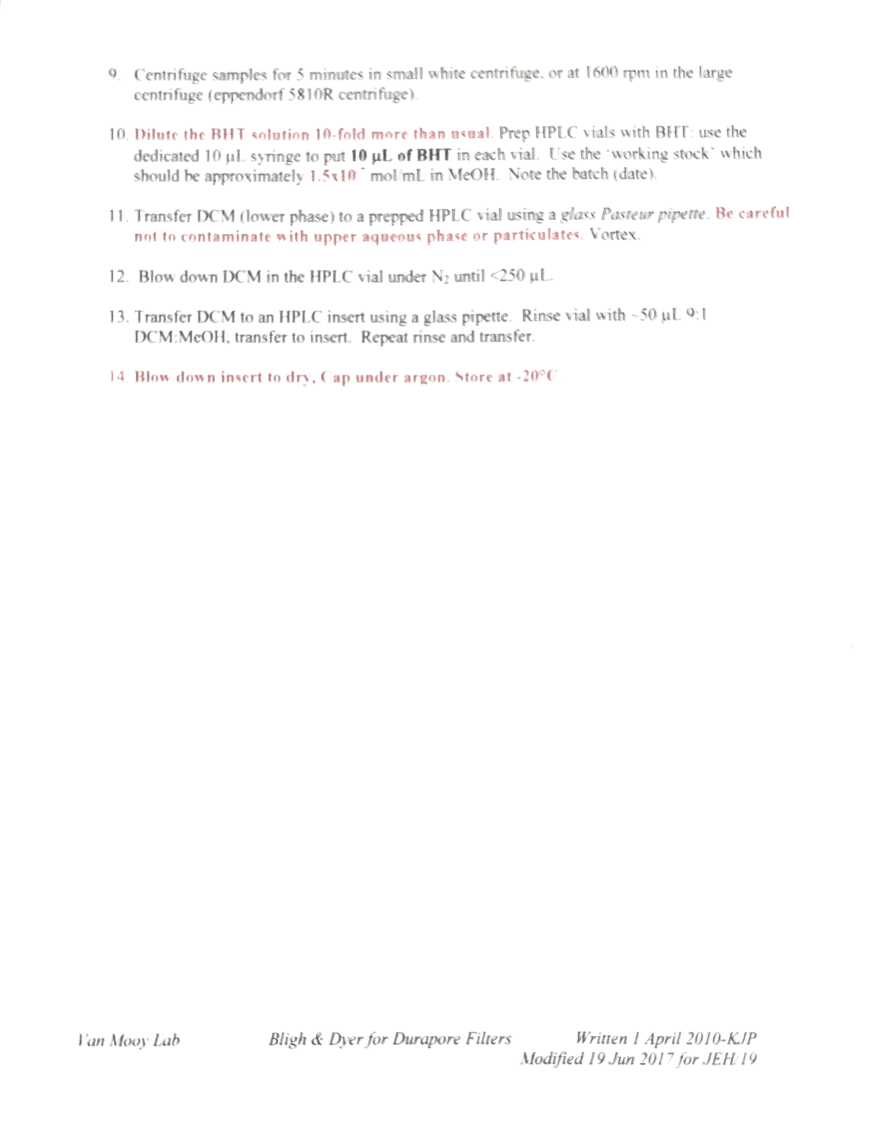
Total lipid extract obtained following a modified Bligh & Dyer protocol. BHT (butylated hydroxyl toluene) added as an antioxidant, DNP-PE added as an internal std (dinitrophenylphosphatidylethanolamine, Avanti polar lipids).
Oxylipin analysis / lipidomics method.
Analyzed on Thermo Exactive plus Orbitrap mass spectrometer (electrospray ionization) coupled to an Agilent 1200 HPLC. Chromatography follows Hummel et al. (2011 Ultra Performance Liquid Chromatography and High Resolution Mass Spectrometry for the Analysis of Plant Lipids. Front. Plant Sci. 2: 54. Doi:10.3389/fpls.2011.00054).
Raw data is presented as .raw files from Xcalibur 2.2 software (Thermo). Conversion utilities are available on the web which will convert .raw files to a more accessible format like .mzXML
Polar membrane lipid analysis
Quantitative: “big 9” lipids on Thermo TSQ Vantage triple quadrupole mass spectrometer. Electrospray ionization, chromatography – normal phase on a diol column as published in Lipids:
Reference: Popendorf, K.J., Fredricks, H.F., and Van Mooy, B.A.S. (2013) Molecular-ion independent quantitation of intact polar diacylglycerolipids in marine plankton using triple quadrupole MS. Lipids 48: 185-195; doi:10.1007/s11745-012-3748-0.
Qualitative: Samples run on Thermo LCQ Fleet. An ion-trap mass spectrometer that provides low-resolution MS and MS/SM data and both positive and negative ion modes. HPLC as above.
Reference: Van Mooy, B.A.S., Fredricks, H.F. (2010). Bacterial and eukaryotic intact polar lipids in the eastern subtropical South Pacific: Water-column distribution, planktonic sources, and fatty acid composition. Geochimica et Cosmochimica Acta, 74 (22): 6499-6516; DOI: 10.1016/j.gca.2010.08.026.
Data
Data custodians:
Experiments 1-5 Helen Fredricks (hfredricks@whoi.edu)
Experiments 6-7 Bethanie Edwards (bedwards@whoi.edu)
Ftp link to raw data files: ftp://ftp.whoi.edu/pub/science/MCG/gbmf/VanMooy/OxylipinAnalysis
Sample list, experiments 1-5 (Excel file)
Sample list, experiments 6 & 7 (Excel file)
1. Diatom cultures exposed to H2O2
Oxylipin analysis: Samples from: Kim Thamatrakoln / Kay Bidle (Rutgers), extracted and analyzed by Helen Fredricks. 5 cultures: T pseudonana, C socialis, C tenuissimus 2-10, C tenuissimus 2-6, C lorenzianus: Each exposed to 0, 30, 150 μM H2O2
2. Pheaodactylum tricornutum culture exposed to H2O2
Oxylipin analysis: Samples from Assaf Vardi group, Weizmann Institute of Plant Sciences, extracted by Jeremy Tagliaferre, analyzed and processed by Helen Fredricks.
Phaeodactylum tricornutum culture exposed to H2O2 at 0, 30 and 150 uM concentrations, samples collected at 4, 8 and 24 hour intervals. In duplicate.
3. Chaetoceros infected with DNA and RNA viruses
Lipid analysis: Samples from: Kim Thamatrakoln / Kay Bidle (Rutgers), extracted by Jeremy Tagliaferre, analyzed and processed by Helen Fredricks. All samples were analyzed on the LCQ, a single replicate from each day/treatment was analysed on the Exactive to give high res. data.
Chaetoceros strains 2-6 and 2-10 exposed to a DNA and a RNA virus. Samples for lipid analysis taken at ~ 1, 3, 5, 7 days. More details in the “Chaetoceros” sheets of the ‘sample list’ excel file.
4. Emiliania Huxleyi strains grown at different calcium concentrations
Lipid analysis. Samples from Chris Johns / Kay Bidle, Rutgers. Samples extracted by Jeremy Taglaferre / Oliver Newman analysed and processed by Helen Fredricks.
Emiliania huxleyi strains 374, 607, 611, 624 and 659 grown at 0.1 and 10 mMol calcium. LCQ / ion trap data files provide low res. data.
5. Viral infection of Emiliania huxleyi
A post-doc in Rutger’s Bidle lab group, Jozef Nissimov has made a couple of visits to the Van Mooy lab to extract and run samples. Samples analyzed by Helen Fredricks and Jozef Nissimov. LCQ ion trap samples provide low resolution MS and MS/MS data in positive and negative ion modes.
6. PS1312- Research cruise in June/July 2013 in the upwelling region of coastal California
a. Water Column Oxylipin Distribution
Particulate Lipid samples were collected by filtering 1L of seawater from 6 depth at 8 stations onto a 0.2 um Durapore filter. Lipids were extracted from the filters back in the lab using a modified Bligh and Dyer (1959) protocol (Popendorf et al. 2013). DNP-PE was added as an internal standard during extraction. Samples for dissolved lipids were collected at 2 depths (Chl max and the 55% PAR) at 8 stations along the cruise track by pre-filtering SW through a 0.2 um sterivex filter to remove particulates. Benzaldehyde was added to the filtrate as an internal standard (10uM). Then, approximately 200ml of filtrate were extracted onto a solid phase extraction (SPE) cartridge (Waters HBL). SPE cartridges were stored at -80C until elution and mass spec analysis (Edwards, in prep). Samples were analyzed using a two different reverse phase HPLC methods paired with high resolution, accurate mass (HRAM) data from a Thermo Q Exactive mass spectrometer (<2 ppm) adapted from Hummel et al. (2011). For quantification of free fatty acids, oxylipins, and intact polar lipid, electrospray ionization-HRAM was paired with a reverse phase chromatographic method using a C8 column (Agilent) as the stationary phase and a starting gradient of 55% water 45% 70:30 ACN:IPA increasing to 1% water 99% 70:30 ACN:IPA over 26 minutes with a 5 min equilibrations period. To optimize for the quantification of polyunsaturated aldehydes, atmospheric pressure chemical ionization-HRAM was paired with a reverse phase chromatographic method using a C18 column (Agilent) as the stationary phase and starting gradient of 80% water 20% 70:30 Methanol:IPA increasing to 1% water 99% 70:30 Methanol:IPA over 26 minutes with a 10 min equilibration period.
b. Oxylipin Distributions in Sinking Particles
From the four traps deployed over 6-12 hours at 50m depth, particulate lipid samples were collected by filtering one-500 ml split of trap material onto a 0.2um Durapore filter. Lipids were extracted from the filters back in the lab using a modified Bligh and Dyer protocol (Popendorf et al. 2013). DNP-PE was added as an internal standard during extraction. Samples for dissolved lipids were collected from the four net traps as well by pre-filtering SW through a 0.2 um sterivex filter to remove particulates. Benzaldehyde was added to the filtrate as an internal standard (10uM). Then, approximately 200ml of filtrate were extracted onto a solid phase extraction (SPE) cartridge (Waters HBL). SPE cartridges were stored at -80C until elution and mass spec analysis (Edwards, in prep). Samples were analyzed using a two different reverse phase HPLC methods paired with high resolution, accurate mass (HRAM) data from a Thermo Q Exactive mass spectrometer (<2 ppm) adapted from Hummel et al. (2011). For quantification of free fatty acids, oxylipins, and intact polar lipid, electrospray ionization-HRAM was paired with a reverse phase chromatographic method using a C8 column (Agilent) as the stationary phase and a starting gradient of 55% water 45% 70:30 ACN:IPA increasing to 1% water 99% 70:30 ACN:IPA over 26 minutes with a 5 min equilibrations period. To optimize for the quantification of polyunsaturated aldehydes, atmospheric pressure chemical ionization-HRAM was paired with a reverse phase chromatographic method using a C18 column (Agilent) as the stationary phase and starting gradient of 80% water 20% 70:30 Methanol:IPA increasing to 1% water 99% 70:30 Methanol:IPA over 26 minutes with a 10 min equilibration period.
c. Oxylipin Incubation Experiments
On-deck incubation experiments were conducted by incubating 20L of whole seawater collected from the 55% PAR depth horizon in the presence of various oxylipin compounds at a range of concentrations in triplicate (see Table ). The purpose of the experiment was to determine the response of natural surface ocean free-living microbial communities to various oxylipins and oxylipin concentrations. After 24 hours of incubation at 55% PAR and in situ temperature the triplicate incubations were harvested. Samples were collected for enzyme activity, particulate and dissolved lipidome, biomass, BSi, dSi, SRP, silicification rates, and viral counts.
| Experiment | Treatment # | Treatment |
| I1 | 1-3 | Control |
| I1 | 4-6 | 0.1 µM PUA |
| I1 | 7-9 | 1 µM PUA |
| I1 | 10-12 | 10 µM PUA |
| I2 | 1-3 | Control |
| I2 | 4-6 | 0.1 µM ARA mix |
| I2 | 7-9 | 1 µM ARA mix |
| I2 | 10-12 | 10 µM ARA mix |
| I3 | 1-3 | Control |
| I3 | 4-6 | 0.1 µM ARA mix |
| I3 | 7-9 | 1 µM ARA mix |
| PUA= heptadienal, octadienal, and decadienal ARA mix= 70% Arachidonic acid, 30% hydroperoxy-eicosatetraenoic acid |
||
Particulate Lipid samples were collected by filtering 1L of seawater from each triplicate onto a 0.2 um Durapore filter. Lipids were extracted from the filters back in the lab using a modified Bligh and Dyer (1959) protocol (Popendorf et al. 2013). DNP-PE was added as an internal standard during extraction. Samples for dissolved lipids were collected by pre-filtering 1 L of each triplicate through a 0.2 um sterivex filter to remove particulates. Benzaldehyde was added to the filtrate as an internal standard (10uM). Then, approximately 200ml of filtrate were extracted onto a solid phase extraction (SPE) cartridge (Waters HBL). SPE cartridges were stored at -80C until elution and mass spec analysis (Edwards, in prep). Samples were analyzed using a two different reverse phase HPLC methods paired with high resolution, accurate mass (HRAM) data from a Thermo Q Exactive mass spectrometer (<2 ppm) adapted from Hummel et al. (2011). For quantification of free fatty acids, oxylipins, and intact polar lipid, electrospray ionization-HRAM was paired with a reverse phase chromatographic method using a C8 column (Agilent) as the stationary phase and a starting gradient of 55% water 45% 70:30 ACN:IPA increasing to 1% water 99% 70:30 ACN:IPA over 26 minutes with a 5 min equilibrations period. To optimize for the quantification of polyunsaturated aldehydes, atmospheric pressure chemical ionization-HRAM was paired with a reverse phase chromatographic method using a C18 column (Agilent) as the stationary phase and starting gradient of 80% water 20% 70:30 Methanol:IPA increasing to 1% water 99% 70:30 Methanol:IPA over 26 minutes with a 10 min equilibration period.
d. Nutrient Amendment experiment
An on-deck nutrient amendment incubation experiment was conducted to determine how the oxylipin profile of natural phytoplankton populations changes under nutrient stress. Whole seawater was collected from the 55% PAR depth horizon and incubated in triplicate under three different conditions: Control (ambient nutrient stress), +NP (simulated Si stress), and +NPSi (replete). After a 72 hour incubation period, samples were collected for the dissolved and particulate lipidome, biomass, enzyme activity, and nutrients.
| Treatment # | Treatment | Nutrient State |
| N1-N3 | Control | Ambient nutrient stress |
| N4-N6 | +NP | Stimulated Si stress |
| N7-N9 | +NPSi | Replete |
Particulate Lipid samples were collected by filtering 1L of seawater from each triplicate onto a 0.2 um Durapore filter. Lipids were extracted from the filters back in the lab using a modified Bligh and Dyer (1959) protocol (Popendorf et al. 2013). DNP-PE was added as an internal standard during extraction. Samples for dissolved lipids were collected by pre-filtering 1 L of each triplicate through a 0.2 um sterivex filter to remove particulates. Benzaldehyde was added to the filtrate as an internal standard (10uM). Then, approximately 200ml of filtrate were extracted onto a solid phase extraction (SPE) cartridge (Waters HBL). SPE cartridges were stored at -80C until elution and mass spec analysis (Edwards, in prep). Samples were analyzed using a two different reverse phase HPLC methods paired with high resolution, accurate mass (HRAM) data from a Thermo Q Exactive mass spectrometer (<2 ppm) adapted from Hummel et al. (2011). For quantification of free fatty acids, oxylipins, and intact polar lipid, electrospray ionization-HRAM was paired with a reverse phase chromatographic method using a C8 column (Agilent) as the stationary phase and a starting gradient of 55% water 45% 70:30 ACN:IPA increasing to 1% water 99% 70:30 ACN:IPA over 26 minutes with a 5 min equilibrations period. To optimize for the quantification of polyunsaturated aldehydes, atmospheric pressure chemical ionization-HRAM was paired with a reverse phase chromatographic method using a C18 column (Agilent) as the stationary phase and starting gradient of 80% water 20% 70:30 Methanol:IPA increasing to 1% water 99% 70:30 Methanol:IPA over 26 minutes with a 10 min equilibration period.
7. Collaborations with Matt Johnson
a. Impacts of growth phase and Si stress on the lipidome of diatom cultures
Culture experiments were conducted to determine how the lipidome of various diatoms shifts with growth phase and Si stress. The particulate and dissolved lipidomes were sampled from cultures of three diatoms isolated from the PS1312 (MJSUR 06, MJSUR12, MJSUR15) and three model diatom species (Chaetoceros socialis, Phaeodactylum tricornutum and Thalassiosira pseudonana) during exponential growth, stationary phase, and Si-limitation (except Pt which does not have a Si requirement).
Particulate Lipid samples were collected by filtering 200 mL of seawater from each triplicate onto a 0.2 um Durapore filter. Lipids were extracted from the filters back in the lab using a modified Bligh and Dyer (1959) protocol (Popendorf et al. 2013). DNP-PE was added as an internal standard during extraction. Samples for dissolved lipids were collected by pre-filtering 200 mL of each triplicate through a 0.2 um sterivex filter to remove particulates. Benzaldehyde was added to the filtrate as an internal standard (10uM). Then, the 200ml filtrate was extracted onto a solid phase extraction (SPE) cartridge (Waters HBL). SPE cartridges were stored at -80C until elution and mass spec analysis (Edwards, in prep). Samples were analyzed using a two different reverse phase HPLC methods paired with high resolution, accurate mass (HRAM) data from a Thermo Q Exactive mass spectrometer (<2 ppm) adapted from Hummel et al. (2011). For quantification of free fatty acids, oxylipins, and intact polar lipid, electrospray ionization-HRAM was paired with a reverse phase chromatographic method using a C8 column (Agilent) as the stationary phase and a starting gradient of 55% water 45% 70:30 ACN:IPA increasing to 1% water 99% 70:30 ACN:IPA over 26 minutes with a 5 min equilibrations period. To optimize for the quantification of polyunsaturated aldehydes, atmospheric pressure chemical ionization-HRAM was paired with a reverse phase chromatographic method using a C18 column (Agilent) as the stationary phase and starting gradient of 80% water 20% 70:30 Methanol:IPA increasing to 1% water 99% 70:30 Methanol:IPA over 26 minutes with a 10 min equilibration period.
b. PtNOA grazing experiment
Samples were collected for complete lipidome analysis from WT Phaeodactylum tricornutum and PtNOA cultures with and without the grazer Oxhyrris marinaat t=2 hrs and t=final over a six hour grazing experiment. The purpose of the experiment was to determine how the lipidome changes under general stress vs. under grazing pressure. Particulate lipid samples were collected by filtering 200 ml of each treatment onto 0.2 µm Durapore filters. The filters were stored at -80˚C and extracted in the lab using a modified Bligh and Dyer (1959) lipid extraction protocol (Popendorf et al. 2013). DNP-PE was added as an internal standard during extraction. Samples for dissolved lipids were pre-filtered through a 0.2 um sterivex filter to remove particulates. Benzaldehyde was added to the filtrate as an internal standard (10uM). Dissolved lipids were then extracted onto a solid phase extraction cartridge (Waters HBL) and stored at -80C until elution and mass spec analysis (Edwards, in prep). Samples were analyzed using a two different reverse phase HPLC methods paired with high resolution, accurate mass (HRAM) data from a Thermo Q Exactive mass spectrometer (<2 ppm) adapted from Hummel et al. (2011). For quantification of free fatty acids, oxylipins, and intact polar lipid, electrospray ionization-HRAM was paired with a reverse phase chromatographic method using a C8 column (Agilent) as the stationary phase and a starting gradient of 55% water 45% 70:30 ACN:IPA increasing to 1% water 99% 70:30 ACN:IPA over 26 minutes with a 5 min equilibrations period. To optimize for the quantification of polyunsaturated aldehydes, atmospheric pressure chemical ionization-HRAM was paired with a reverse phase chromatographic method using a C18 column (Agilent) as the stationary phase and starting gradient of 80% water 20% 70:30 Methanol:IPA increasing to 1% water 99% 70:30 Methanol:IPA over 26 minutes with a 10 min equilibration period.
Next Step: Lipidome Annotation
The particulate and dissolved lipidomes are now being annotated in the software package, Metabolic Analysis and Visualization ENgine (MAVEN,) that detects peaks and facilitates pseudo-targeted lipidomics analysis (Melamud et al., 2010; Collins et al. in prep). Identities are assigned to individual peaks by querying a lipid database that Helen Fredricks populated with IPLs, FA, oxylipins, pigments, and sterols. The ability to assign ID based on m/z hinges on the high mass resolution afforded to us by the Thermo Q Exactive mass spec.
Works cited:
Bligh EG, Dyer WJ (1959) A rapid method of total lipid extraction and purification. Can. J. Physiol. Pharmacol. 37:911–917.
Collins JR et al. in prep. Pseudo-targeted lipidomics analysis of cultured marine microbes and natural marine microbial communities.
Edwards BR et al. in prep. A Novel Method for the Extraction and Quantification of Particulate and Dissolved Oxylipins from Marine Environments.
Hummel J et al. (2011) Ultra Performance Liquid Chromatotraphy and High Resolution Mass Spectrometry for the Analysis of Plant Lipids. Front. Plant Sci. 2: 54. Doi:10.3389/fpls.2011.00054
Melamud E et al. (2010) Metabolomic Analysis and Visualization Engine for LC-MS Data. Anal. Chem. 82: 9818-9826. Doi: 10.1021/ac1021166
Popendorf KJ et al. (2013) Molecular Ion-Independent Quantification of Polar Glycerolipid Classes in Marine Plankton Using Triple Quadrupole MS. Lipids 48:185-195. Doi:10.1007/s11745-012-3748-0
Related Files:
» Sample list, experiments 1-5
» Sample list, experiments 6 & 7
Related Links» Oxylipin Analysis raw data files
E. Huxleyi haploid / diploid + virus
Van Mooy Lab data files: Emiliania huxleyi (haploid & diploid strains) subject to virus EhV201.
The experiment and data are best described in the following manuscript – currently awaiting publication in Frontiers in Marine Science section Aquatic Microbiology (as of 9/25/15). Title and abstract below. For further information please contact data custodian Helen Fredricks at hfredricks@whoi.edu.
Targeted and Untargeted Lipidomics of Emiliania huxleyi Viral Infection and Life Cycle Phases Highlights Molecular Biomarkers of Infection, Susceptibility, and Ploidy.
Jonathan E. Hunter†1,2, Miguel J. Frada3, Helen F. Fredricks4, Assaf Vardi3 and Benjamin Van Mooy4.
1. Ocean & Earth Science, University of Southampton, National Oceanography Centre, European Way, Southampton, SO14 3ZH, United Kingdom
2. Institute for Life Sciences, University of Southampton, SO17 1BJ, United Kingdom
3. Department of Plant and Environmental Sciences, Weizmann Institute of Science, Rehovot 76100, Israel
4. Department of Marine Chemistry and Geochemistry, Woods Hole Oceanographic Institution, Woods Hole, Massachusetts, 02543, United States of America.
†Corresponding Author: J.Hunter@soton.ac.uk; Ocean and Earth Science, University of Southampton, National Oceanography Centre, European Way, SO14 3ZH, United Kingdom.
Abstract
Marine viruses that infect phytoplankton strongly influence the ecology and evolution of their hosts. Emiliania huxleyi is characterized by a biphasic life cycle composed of a diploid (2N) and haploid (1N) phase; diploid cells are susceptible to infection by specific coccolithoviruses, yet haploid cells are resistant. Glycosphingolipids (GSLs) play a role during infection, but their molecular distribution in haploid cells is unknown. We present mass spectrometric analyses of lipids from cultures of uninfected diploid, infected diploid, and uninfected haploid E. huxleyi. Known viral GSLs were present in the infected diploid cultures as expected, but surprisingly, trace amounts of viral GSLs were also detected in the uninfected haploid cells. Sialic-acid GSLs have been linked to viral susceptibility in diploid cells, but were found to be absent in the haploid cultures, suggesting a mechanism of haploid resistance to infection. Additional untargeted high-resolution mass spectrometry data processed via multivariate analysis unveiled a number of novel biomarkers of infected, non-infected, and haploid cells. These data expand our understanding on the dynamics of lipid metabolism during E. huxleyi host/virus interactions and highlight potential novel biomarkers for infection, susceptibility, and ploidy.
Polar membrane lipid analysis (LCQ) / lipidomics analysis (Exactive)
Sample: Total lipid extract obtained following a modified Bligh & Dyer protocol. BHT (butylated hydroxyl toluene) added as an antioxidant, DNP-PE added as an internal std (dinitrophenylphosphatidylethanolamine, Avanti polar lipids).
LCQ data: Analyzed on Thermo LCQ Fleet ion-trap mass spectrometer (electrospray ionization) coupled to an Agilent 1100 HPLC. Chromatography follows Popendorf et al: Popendorf, K.J., Fredricks, H.F., and Van Mooy, B.A.S. (2013) Molecular-ion independent quantitation of intact polar diacylglycerolipids in marine plankton using triple quadrupole MS. Lipids 48: 185-195; doi:10.1007/s11745-012-3748-0.
Exactive Plus data: Analyzed on Thermo Exactive plus Orbitrap mass spectrometer (electrospray ionization) coupled to an Agilent 1200 HPLC. Chromatography follows Hummel et al: 2011 Ultra Performance Liquid Chromatography and High Resolution Mass Spectrometry for the Analysis of Plant Lipids. Front. Plant Sci. 2: 54. doi:10.3389/fpls.2011.00054.
Raw data is presented as .raw files from Xcalibur 2.2 software (ThermoScientific). Conversion utilities are available on the web which will convert .raw files to a more accessible format like .mzXML
Related Files:» GBMF data haploid diploid E Hux
Related Links:» E. Huxleyi haploid / diploid + virus data